Disease
Coral Disease
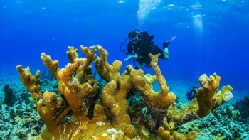
The Study of Coral Diseases
Coral diseases are conditions, within a coral, that impair the biotic functions which allow the coral systems to thrive. These diseases are caused by external, internal, biological, and non-biological stressors. It is thought that a combination of stressors is often the cause of disease. [2]
As the study of coral disease is a relatively new field, many obstacles stand in the way of obtaining the desired knowledge on the subject. One of these obstacles is the variety of diseases that have been discovered. Within each category of coral disease, there are multiple types that can be identified. This makes research on the topic increasingly complex. A further complication is the lack of knowledge concerning the causes of coral disease. Due to this, it is challenging to find treatments for these diseases. [3] However, the temporal and spatial distributions of coral diseases are known to predict impacts of disease, knowing this helps current reef management prevent the spread and severity of the diseases. [4]
Current research is also using management studies, legislation, and new technologies to mitigate the continued spread of coral disease. Over the past decade, diseases affecting coral reefs have increased in frequency and severity. [5] The impacts of this increase have been exceptionally significant in the Caribbean, where up to 80% of the coral has been decimated. [6]
The Stressors
Non-Biological

The three main non-biological stressors believed to cause coral diseases are increased sea surface temperature (SST), increased ultraviolet (UV) radiation, and pollution. [2] Of these, the most concerning is increased SST. Many studies have indicated that the current trend of global temperature increase could result in SST increasing by one degree in the next 50 years. An increase such as this would cause local climates all over the globe to shift, thus severely impacting coral ecosystems. The symbiotic relationships with bacteria that sustain coral are predicted to break down completely if this occurs.
Along with increased SST, increased UV radiation is also a concern. If UV radiation continues to increase around the world, the problems caused by increased SST would only worsen. Larger amounts of UV radiation reaching coral reefs also brings its own problems, such as decreased autotrophic capacity, grazing rates, and calcification rates within the coral colonies. Although these are not direct causes of disease, these effects would weaken coral colonies and put them at higher risk of infection. [8]
The issue of pollution brings more complications for coral colonies despite being synthetic or natural. Pollution alone can smother corals and prevent them from continuing to thrive. Pollution that drifts onto reefs may break off sections of coral, or alter essential structures on the reefs. It can also lead to major food structure alterations that negatively impact corals. [9]
However, pollution can harm coral reef systems without coming into contact with them as well. Water quality often decreases due to the presence of pollution. As the quality decreases, the growth rates of harmful algae often increase on reefs, again putting the coral at higher risk for disease.[10]
Biological
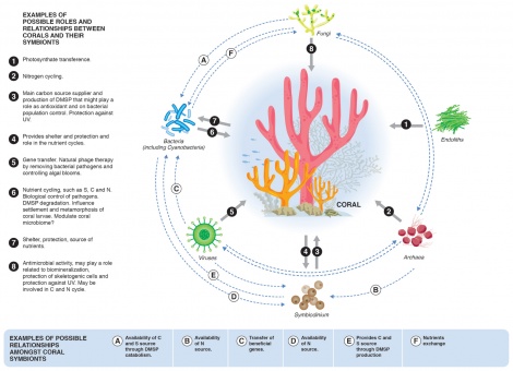
Biological stressors are caused by changes in the naturally occurring bacteria and fungi, which are usually helpful, that exist in coral reef ecosystems. In conjunction with non-biological stressors, these systems can become unbalanced, resulting in too many bacteria and fungi or new and harmful types of bacteria and fungi. When this occurs, the microorganisms can attack the corals; this leads to increased susceptibility to disease. [11]
Main Coral Diseases in the Caribbean
Dark Spots Disease
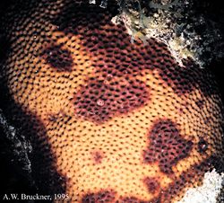
Dark spots syndrome is seen by dark gray, purple, or brown spots scattered on the coral surface that increase in size and radiate outward as the affected area dies. [14] The individual, darkened polyps are often depressed and smaller in size. There is no known pathogen for DSS and interestingly, seawater temperatures do not seem to correlate with the percent of DSS cover in a study done by Dr. Deborah Gochfeld on massive starlet coral. The study discovered 2 to 4 strains of ecologically relevant bacteria in which the coral had demonstrated antimicrobial activity. [15]This shows the corals do possess chemical defenses to bacteria, but scientists are not aware as of to which ones. Some bacteria may be harmful, beneficial, or inconsequential. The study concluded a lot of temporal and spatial variation - one reason suggests that this is due to microbial community variability. In advanced cases of DSS, the skeleton of the coral may erode, leaving an indentation in the coral that is usually filled by algae. [16]
Black Band Disease
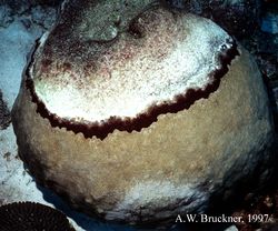
Black band disease was the first described coral disease. It is characterized by a blackish concentric or crescent shaped band, also known as a microbial mat. This microbial mat is made up of photosynthetic and non-photosynthetic bacteria that co-exist: the three functionally and physically dominant bacteria are cyanobacteria, sulfate reducing, and sulfide-oxidizing. Cyanobacteria gives the photosynthetic pigment of the black band. The sulfur bacteria create white specks. Live coral tissue is consumed as the microbial mat passes over the colony surface, leaving behind fully exposed coral skeletons. The microbial mat moves at rates of 3mm to 1cm a day. The tissue death is caused by exposure to an anoxic, sulfide-rich microenvironment associated with the base of the band. The only known reservoir is within cyanobacterial biofilms. [17]
Red band disease
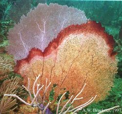
Two types of red band disease exist: RBD-1, which closely resembles black band disease, except that the bands are maroon in color and lacks the white specks; RBD-2 has cyanobacterial filaments that spread like a net on the surface. RBD leaves behind exposed coral skeleton. The pigment is caused by a different genus of cyanobacteria as the dominant microorganism, and the migration pattern across the surface of the colony also differs from BBD.[18]
White Band Disease
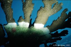
White band disease is a common coral disease detrimental to elkhorn and staghorn coral. [14] Affected corals have thick strips of bleaching occurring at the bottom of the branch that spread upwards. The bleaching is caused by the expulsion of symbiotic algae while tissue pulls away from the skeleton starting at the base. It is suspected that WBD is caused by a bacterial infection. [19] There are two types of the disease, White Band Disease 1 (WBD1) and White Band Disease 2 (WBD2). The spread of the disease must be observed in order to determine which type it is. In type 1 there is a strip of lysis (cell break down), and in type 2 the coral degradation eventually catches up to this line of lysis.
This is one of the only diseases known to cause major changes in the structural composition of reefs. This disease was first documented in 1979, and since that time it has destroyed up to 95% of the acropora genus corals in the Caribbean.[20]
White Plague Disease
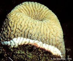
White plague disease is similar to the white band disease in that it causes portions of coral to bleach. However, the white plague disease affects a wider range of coral. A symptom of this disease is irregular lesions that leave white patches on coral. [14] White plague disease has a similar pattern to the WBD that the tissue begins to pull away from the bottom leaving a large line of bleached coral.
Yellow Blotch Disease
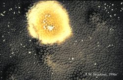
Yellow blotch disease causes sections of corals to become yellowed and translucent. Like many other coral diseases, the cause is unknown.[21] The disease begins at a central point anywhere on the coral and spreads outward. This is caused by a bacterial infection that causes yellowing of the tissue. As the infection spreads, the tissue is still alive, but eventually the tissue at the central point begins to die. This disease mostly affects large boulder star corals. Unlike most other diseases, recently bleached, white skeleton is rarely observed; typically the area behind the band is covered in filamentous and coralline algae.
Aspergillosis
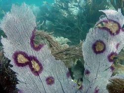
Aspergillosis, caused by the Aspergillus sydowii fungus, primarily affects the Caribbean sea fan and other gorgonian corals. The disease ranges from severe to mild, with the ability to cause partial tissue loss or mass localized mortalities. In most cases, symptoms develop only a few days after infections. [23] In Gorgonia ventalina, or purple sea fans, aspergillosis results in damaged areas and spots that can be identified by a deep purple color. [24]
Labyrinthulomycetes Microbes
Purple sea fans are also affected by Aplanochytrium, a member of an order of lethal microbes known as Labyrinthulomycetes. Aplanochytrium grows faster at warmer temperatures, so there is concern that this pathogen could play a larger role with climate change. [25]
Stony Coral Tissue Loss Disease (SCTLD)
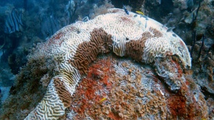
Stony coral tissue loss disease, or SCTLD, is a relatively new coral disease, first reported in Florida in 2014. [27] Currently, SCTLD is the cause of significant damage on reefs in Florida and the Caribbean Islands. [28] Outbreaks of SCTLD have been reported in parts of the Caribbean, such as Jamaica, Mexican Caribbean, St. Maarten, USVI, the Dominican Republic, and most recently, St. Thomas. [27]
There is a great concern for Caribbean reefs, specifically, as SCTLD has an extensive geographic and temporal range. The disease also affects many different species of coral and has high mortality rates. Bacterial Pathogens are the suspected cause of SCTLD, as researchers have discovered that the disease can be transmitted via direct contact and water circulation. [28]
Implications of Diseases in Reefs
Coral reefs are an important resource for coastal communities. Reefs are important for economies since they bring in money through tourism and the fishing industry. The weakening reef system may pose a threat to the countries that rely on this ecosystem as a major component of their economy. Diseased reefs do not have the same tourist draw or fishing production as a healthy reef. Furthermore, reefs provide a barrier to protect the land from large waves and surges. [29] Coral disease outbreaks have major negative implications. Therefore, more work and research should focus on the causes in order to prevent further degradation of the reef ecosystem.
Coral disease hasn’t been thoroughly studied because it does present some challenges. First, it is hard to run experiments on sick corals that may have very short life spans and migrates across the coral. Second, there is an enormous variety of microbes and bacteria that cause disease. Lastly, spatial and temporal variation adds an exponential amount of variability when trying to generalize results. We need more marine microbiologists who can conduct research in the field and also understand the ecological and environmental effects on coral.
We must also consider that even though the diseases can very much occur naturally, humans can and do impose threats that may spread or worsen the toll disease takes on corals. Pollution and waste may be facilitators in spreading disease. Sea rise and temperature increase may increase the susceptibility of coral, and ocean acidification may weaken the corals’ immune systems. It is important that we do our part to protect the reef. By making small changes and encouraging sustainable behavior within the tourism industry, it can help the health of the reef ecosystem. Many of the chemicals used in sunscreen and recreational activities (golfing, boating, etc.) can cause added stress to corals, which make them more susceptible to diseases.
Coral Disease Remediation
Developing a Coral Disease Response Plan
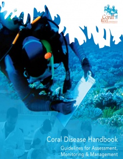
A coral disease response plan “describes the steps for detecting, assessing, and responding to disease outbreaks.” [31]Such plans include pin-pointing affected areas, finding funding for responsive projects, projecting which areas are most susceptible to outbreaks in the future, and planning how to maintain a response plan for the duration of the disease. Making the formation of a plan (such as the one detailed by Raymundo et. al (2008)) [30] is the best way to ensure projects are able to maintain themselves for the duration of the outbreak.
In the Great Barrier Reef, scientists have been successful in creating a method to locate the most vulnerable reefs. [32] Their study concentrates on White Syndromes, Black Band and Brown Band disease with the primary objectives of hindcasting where outbreaks are likely to have occurred and forecasting where outbreak likelihood would be high, and subsequently surveying these reefs to improve the knowledge of conditions triggering outbreaks. This information will help to concentrate efforts where they are needed most in such a large area, as well as to determine the best management responses. Climate change projections suggest that the conditions that caused the 2002 Great Barrier Reef outbreaks and the White Syndrome outbreak of 2009 will occur more frequently in the future. The method used by Maynard et. al[32] highlights that an increased frequency and severity of bleaching events in the future could increase disease susceptibility and lower the coral immune health threshold required for outbreaks. Developing new tools and refining existing ones such as those applied in the Great Barrier Reef will be critical in increasing our understanding of outbreak causation and capacity to defend our reefs in the face of climate change. After the affected and vulnerable areas have been determined, direct action and policy tools may be applied to halt the spread of the disease.
Direct Management
Direct management includes any response that involves hands-on action. Examples include aspirating diseased corals with syringes or pumps or surgically removing diseased portions. Syringes and pumps have been successful with black band disease, yellow band disease, white band disease, and white plague.[31] With direct management actions such as these, there is a significant risk of spreading the disease in the process.[31] These direct measures are not a means of completely eradicating the disease, but may be a viable approach to save certain high-value colonies, such as massive reef building corals, or rare species that are viewed as particularly important as well as slowing the spread of the disease. Additionally, such responses adhere immediate results (though potentially not long lasting).
Policy Tools
A coral's immune system may be lowered by sedimentation, water quality, bleaching, or translocation. [31] Many of these factors are exacerbated by humans, and thus policies can be implemented to reduce human impact. Policies may be set in place that reduce or restrict human access to reefs to reduce translocation and sedimentation, such as limiting or eliminating diving and fishing. Additionally, policies may restrict the take of herbivorous fish so that algal grazers can keep algal blooms under control.[31] Such measures (when tailored towards factors triggering disease) can be very effective in limiting the way humans increase the spread of disease.
Restoration Methods
Quarantine Healthy Corals
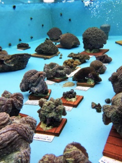
In Florida, scientists are “rescuing” healthy corals from a disease outbreak by moving them into tanks, isolating the corals from future spread of disease. The study is focused on Stony Coral Tissue Loss disease in the St. Thomas outbreak. This quarantine of healthy corals allows scientists to study and breed healthy corals, while simultaneously providing a safe way to study the diseased corals without causing further spread. With this combined research, the team is working to create an antibiotic and has seen an 85% success rate. The team hopes to transplant captive corals back into the reef once the outbreak subsides.[33]
Coral Repopulation
Since 2014, the Florida Tract has experienced a disease outbreak of Stony Coral Tissue Loss Disease that has devastated surrounding reefs. As much as 35% of the local coral population has been lost due to this outbreak. [34] The Stony Coral Tissue Loss Disease is a fast acting disease that, once found in a colony, can kill the colony within days, weeks, or months. The disease starts with tissue loss at the base of the coral that starts in a band or patch and extends upward over time.[35] While many organizations are making efforts to mitigate the impacts of this coral disease, Mote laboratories is one facility making leeway in coral reef revival efforts as a result of this outbreak.
Mote Marine Laboratory and Aquarium is an independant, non-profit, marine research institution located in Florida that has a Coral Health & Disease Program that has been studying the causes and consequences of coral disease along with ways to prevent and remedy diseased corals.[36] The laboratory worked with the Division of Aquatic Resources in Hawaii and the Institute of Marine Biology in Kaneohe Hawaii to develop and test the efficiency of a new strategy of coral transplantation to help recover coral populations specifically in response to the recent coral disease outbreak off of Florida’s coast. [34] These relief efforts are especially critical in recent years as disease rates in coral drastically increase with higher water temperatures, such as the white pox disease on the threatened elkhorn coral that was a result of the higher temperatures stressing the hosts. These marine biologists have developed a new technique of transplantation called micro-colony-fusion, which accelerates coral cover and helps impacted coral areas recover faster.[36]
In this technique of micro-colony-fusion, small fragments of coral from the same colony are spread over ceramic tiles to grow, which increases spreading rates and is then followed by isogenic fusion[37]. Fusion had previously been shown to reduce size-specific mortality in juvenile corals. This micro-fragmentation-fusion strategy manipulates the surface area of coral onto a flat surface, which allows the small colonies to spread tissue rapidly and fuse. Over a four-month experimental period, micro-fragments of O. faveolata increased 293 cm2 while P. clivosa fragments grew 222 cm2. [37] The transplantation process yielded a high growth rate in the coral cover of 329% and 154% increase in area respectively over the four months.[37] This process effectively combines the benefits of coral transplantation and that of artificial reefs while speeding up the growth rates of the coral. While more studies about the genotypic, physiological, and reproductive effects of the process are necessary to fully understand the benefits of this new form of transplantation, it effectively lowers the mortality rate for juvenile corals below that of traditional transplantation processes.[37] There are current field trials to further develop methods to efficiently encourage nursery-grown coral to fuse and self-attach over the benthic substrate, which would allow the replenishment of reef areas that have been completely wiped out by disease and other threats.

CRISPR
Scientists have identified the potential for CRISPR/Cas-9 technology to aid in preventing coral disease at a genetic level. CRISPR is an acronym, and it stands for “clusters of regularly interspaced short palindromic repeats.” This refers to nucleotide triplets that code for amino acids, which can have patterns of nucleotide bases. Spacers are the DNA between those points. Cas-9 is a protein designed to cut DNA. It recognizes the sequences of repeated nucleotides as places to start and stop cutting. Research developed in 2012 discovered how to create “guideRNA”, a combination of two other types of RNA that allow it to cut at any space in foreign DNA, which opened up many more possibilities for the technology.[39] Among them was the idea that CRISPR/Cas-9 could be used to slightly alter the genome of coral species and provide immunity to disease, or lessen the potential effects of catching a disease.
A 2016 study on the critical reef-building coral Acropora Millepora tested the use of CRISPR on three genes. This was a test to examine techniques and possibilities with editing coral genomes with the idea that if it works, the strategy can be used to target genes that increase immunity or strength. The researchers targeted two fluorescent proteins and a fibroblast growth factor, which allowed them to determine with certainty whether or not the editing was successful. They detected the desired mutation in ~50% of species grown after editing, which, while not perfect, is far better than 0%. However, 100% of larvae experienced mutations outside of the target gene. They survived and grew, but further research is needed to determine the range and implications of effects after the target gene is edited.[40] Research like this serves to prove that the potential for a positive impact on disease resistance using CRISPR technology is definitely a possibility. However, given the potential risk of harmful mutations, and the overall dearth of research on the topic, it is not in a stage where it could be implemented at the moment.
An important part of using CRISPR technology is understanding which part of the agent’s DNA is allowing it to affect coral, and how it maintains its virulence. In 2016 a study was done that examined the genome of two cyanobacterium associated with Black Band Disease, which contained potential virulence genes and were equipped with a CRISPR-Cas defense system. Researchers were able to identify three potential prophages in one and five in the other.[41] This provides insight as to how Black Band Disease protects itself from bacteriophage predators and how it targets coral, allowing researchers to explore protecting coral by altering the specific sequence that disease affects, thus furthering the field.
With the lack of current research, and very little information on what the scope of preliminary projects would be estimating the cost of using this technology to grow corals and introduce them to the natural environment cannot be accurately estimated, but trends in other fields of science show that the process and development would be exorbitantly expensive. As it stands now, expense and the potential for causing more damage through mutations are two of the biggest drawbacks to using CRISPR/Cas-9 to help increase coral disease resistance.
References
- ↑ https://e360.yale.edu/features/as-disease-ravages-coral-reefs-scientists-scramble-for-solutions/
- ↑ 2.0 2.1 ”Corals” NOAA Ocean Service Education. NOAA, 01-07-20. Web. 02-29-20 <https://oceanservice.noaa.gov/education/kits/corals/coral10_disease.html>
- ↑ https://www.ncbi.nlm.nih.gov/pmc/articles/PMC3197597//
- ↑ https://www.ncbi.nlm.nih.gov/pmc/articles/PMC4473065//
- ↑ http://coraldisease.org/
- ↑ “Coral Diseases” Reef Research Center. Retrieved 26 Feb. 2013 from <http://www.reef.crc.org.au/discover/coralreefs/Coraldisease.htm>
- ↑ https:https://reefresilience.org/stressors/local-stressors/pollution/
- ↑ https://aslopubs.onlinelibrary.wiley.com/doi/pdf/10.1002/lno.10481?casa_token=QQ0zQHXNnmkAAAAA:NBwwNb1a4-hI1GytyTacPi_sOyEkNx8qvTPdSRfBJKRg0nTsC6Mq0u5MvXOE44h-Xrv6CRYXjMRnlA0/
- ↑ https://oceanservice.noaa.gov/education/tutorial_corals/coral09_humanthreats.html/
- ↑ https://floridakeys.noaa.gov/corals/pollution.html/
- ↑ https://link.springer.com/referenceworkentry/10.1007%2F978-3-642-30123-0_48/
- ↑ https://www.frontiersin.org/articles/10.3389/fmicb.2017.00341/full/
- ↑ 13.0 13.1 13.2 13.3 13.4 13.5 Field Guide to Western Atlantic Coral Diseases and Other Causes of Coral Mortality. http://www.masna.org/portals/0/NOAACoralDisease/index.htm
- ↑ 14.0 14.1 14.2 “Major Reef-building Coral Diseases.” CoRIS - Coral Reef Information System. NOAA, 01-17-13. Web. 2-26-13. <http://coris.noaa.gov/about/diseases/#redband>
- ↑ Gochfield, Deborah, Julie Olson, and Marc Slattery. “Colony Versus Population Variation in Susceptibility and Resistance to Dark Spot Syndrome in the Caribbean Coral Siderastrea Siderea.” Diseases of Aquatic Organisms 69 (2006): 53–65. Inter-Research. Web. 02-26-13. <http://www.int-res.com/abstracts/dao/v69/n1/>
- ↑ http://coraldisease.org/diseases/12
- ↑ http://www.artificialreefs.org/Corals/diseasesfiles/Common%20Identified%20Coral%20Diseases.htm#BlackBand
- ↑ http://archive.rubicon-foundation.org/xmlui/handle/123456789/9047
- ↑ Aronson, Richard B. and Precht, William F. “White-band disease and the changing face of Caribbean coral reefs.” 2009. Hydrobiologia 460: 25-38
- ↑ Lentz JA, Blackburn JK, Curtis AJ (2011) Evaluating Patterns of a White-Band Disease (WBD) Outbreak in Acropora palmata Using Spatial Analysis: A Comparison of Transect and Colony Clustering. PLoS ONE 6(7): e21830. doi:10.1371/journal.pone.0021830
- ↑ Kellogg, C. A. “Montastraea cavernosa.” Photo. microbiology.usgs.gov 26 Feb. 2013. <http://microbiology.usgs.gov/image_gallery_plants_animals_montastraea_cavernosa.html>
- ↑ https://www.researchgate.net/figure/Picture-of-a-sea-fan-coral-Gorgonia-ventalina-infected-with-Aspergillus-sydowii_fig11_5487449/
- ↑ https://onlinelibrary.wiley.com/doi/10.1002/9781118828502.ch16/
- ↑ https://www.int-res.com/articles/dao2014/109/d109p257.pdf/
- ↑ http://www.frontiersin.org/Invertebrate_Physiology/10.3389/fphys.2013.00180/abstract
- ↑ https://www.whoi.edu/news-insights/content/virgin-island-corals-in-crisis//
- ↑ 27.0 27.1 "Stony coral tissue loss disease". International Coral Reef Initiative. Accessed May 2019. https://www.icriforum.org/news/2019/04/stony-coral-tissue-loss-disease
- ↑ 28.0 28.1 "Stony Coral Tissue Loss Disease". Reef Resilience Network. RRN, 2020. Web. 03-31-20<http://reefresilience.org/managing-for-disturbance/managing-coral-disease/stony-coral-tissue-loss-disease//
- ↑ “Socioeconomic Impacts” Coral Reefs. Retrieved 26 Feb. 2013 from <http://www.reefresilience.org/Toolkit_Coral/C2c2_Socioecon.html>
- ↑ 30.0 30.1 <https://reefresilience.org/pdf/Raymundo_etal_2008.pdf>
- ↑ 31.0 31.1 31.2 31.3 31.4 <https://reefresilience.org/managing-for-disturbance/managing-coral-disease/ >
- ↑ 32.0 32.1 Maynard, J.A., Anthony, K.R.N., Harvell, C.D. et al. Predicting outbreaks of a climate-driven coral disease in the Great Barrier Reef. Coral Reefs 30, 485–495 (2011)<https://doi-org.libproxy.lib.unc.edu/10.1007/s00338-010-0708-0)>
- ↑ 33.0 33.1 "A mysterious coral disease is ravaging Caribbean reefs" ScienceNews. ScienceNews, 08-03-19. Web. 02-18-20<https://www.sciencenews.org/article/mysterious-coral-disease-ravaging-caribbean-reefs/
- ↑ 34.0 34.1 <https://e360.yale.edu/features/as-disease-ravages-coral-reefs-scientists-scramble-for-solutions>
- ↑ <https://sanctuaries.noaa.gov/news/aug18/coral-disease-mystery-florida-keys.html>
- ↑ 36.0 36.1 <https://mote.org/research/program/coral-health-and-disease>
- ↑ 37.0 37.1 37.2 37.3 <https://peerj.com/articles/1313/>
- ↑ https://www.researchgate.net/figure/CRISPR-Cas9-mediated-gene-editing-mechanisms-A-single-guide-RNA-sgRNA-recognizes-a_fig1_322251194/
- ↑ <https://www.livescience.com/58790-crispr-explained.html>
- ↑ <https://www.pnas.org/content/115/20/5235>
- ↑ <https://www.ncbi.nlm.nih.gov/pmc/articles/PMC5177637/>