Disease: Difference between revisions
BrianNaess (talk | contribs) |
BrianNaess (talk | contribs) |
||
| Line 10: | Line 10: | ||
===Dark Spots Disease=== | ===Dark Spots Disease=== | ||
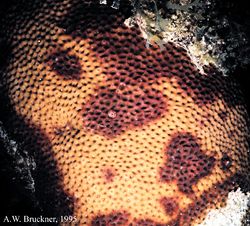
Dark spots syndrome is seen by dark gray, purple, or brown spots scattered on the coral surface that increase in size and radiate outward as the affected area dies. <ref>“Major Reef-building Coral Diseases.” CoRIS - Coral Reef Information System. NOAA, 01-17-13. Web. 2-26-13. <http://coris.noaa.gov/about/diseases/# | Dark spots syndrome is seen by dark gray, purple, or brown spots scattered on the coral surface that increase in size and radiate outward as the affected area dies. <ref name="major">“Major Reef-building Coral Diseases.” CoRIS - Coral Reef Information System. NOAA, 01-17-13. Web. 2-26-13. <http://coris.noaa.gov/about/diseases/#redband></ref> The individual, darkened polyps are often depressed and smaller in size. There is no known pathogen for DSS and interestingly, seawater temperatures do not seem to correlate with the percent of DSS cover in a study done by Dr. Deborah Gochfeld on massive starlet coral. The study discovered 2 to 4 strains of ecologically relevant bacteria in which the coral had demonstrated antimicrobial activity. <ref>Gochfield, Deborah, Julie Olson, and Marc Slattery. “Colony Versus Population Variation in Susceptibility and Resistance to Dark Spot Syndrome in the Caribbean Coral Siderastrea Siderea.” Diseases of Aquatic Organisms 69 (2006): 53–65. Inter-Research. Web. 02-26-13. <http://www.int-res.com/abstracts/dao/v69/n1/></ref>This shows the corals do possess chemical defenses to bacteria, but scientists are not aware as of to which ones. Some bacteria may be harmful, beneficial, or inconsequential. The study concluded a lot of temporal and spatial variation - one reason suggests that this is due to microbial community variability. In advanced cases of DSS, the skeleton of the coral may erode, leaving an indentation in the coral that is usually filled by algae. <ref>http://coraldisease.org/diseases/12</ref> | ||
<hr style="clear:both" /> | <hr style="clear:both" /> | ||
[[File:bbd.jpg|thumb|250x208px|left|Black Band Disease <ref name="msna" />]] | [[File:bbd.jpg|thumb|250x208px|left|Black Band Disease <ref name="msna" />]] | ||
===Black Band Disease=== | ===Black Band Disease=== | ||
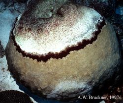
Black band disease was the first described coral disease. It is characterized by a blackish concentric or crescent shaped band, also known as a microbial mat. This microbial mat is made up of photosynthetic and non-photosynthetic bacteria that co-exist: the three functionally and physically dominant bacteria are cyanobacteria, sulfate reducing, and sulfide-oxidizing. Cyanobacteria gives the photosynthetic pigment of the black band. The sulfur bacteria create white specks. Live coral tissue is consumed as the microbial mat passes over the colony surface, leaving behind fully exposed coral skeletons. The microbial mat moves at rates of 3mm to 1cm a day. The tissue death is caused by exposure to an anoxic, sulfide-rich microenvironment associated with the base of the band. The only known reservoir is within cyanobacterial biofilms. <ref>http://www.artificialreefs.org/Corals/diseasesfiles/Common%20Identified%20Coral%20Diseases.htm#BlackBand</ref> | Black band disease was the first described coral disease. It is characterized by a blackish concentric or crescent shaped band, also known as a microbial mat. This microbial mat is made up of photosynthetic and non-photosynthetic bacteria that co-exist: the three functionally and physically dominant bacteria are cyanobacteria, sulfate reducing, and sulfide-oxidizing. Cyanobacteria gives the photosynthetic pigment of the black band. The sulfur bacteria create white specks. Live coral tissue is consumed as the microbial mat passes over the colony surface, leaving behind fully exposed coral skeletons. The microbial mat moves at rates of 3mm to 1cm a day. The tissue death is caused by exposure to an anoxic, sulfide-rich microenvironment associated with the base of the band. The only known reservoir is within cyanobacterial biofilms. <ref>http://www.artificialreefs.org/Corals/diseasesfiles/Common%20Identified%20Coral%20Diseases.htm#BlackBand</ref> | ||
Revision as of 10:28, 21 May 2013
Coral Disease
Why Study Coral Diseases?
Diseases affecting coral reefs have increased in frequency and severity in recent decades. [1] The impact has been exceptionally significant in the Caribbean in which up to 80% of the coral has been decimated. [2] One of the major problems of coral diseases is that there are a variety of diseases, and within each, there are multiple types. Further complicating the situation, many of the causes of the diseases are unknown, which makes it difficult to treat. The temporal and spatial distributions of coral disease predict impacts that help current reef management prevent the spread and severity of disease.
Main Coral Diseases in the Caribbean
Dark Spots Disease
Dark spots syndrome is seen by dark gray, purple, or brown spots scattered on the coral surface that increase in size and radiate outward as the affected area dies. [4] The individual, darkened polyps are often depressed and smaller in size. There is no known pathogen for DSS and interestingly, seawater temperatures do not seem to correlate with the percent of DSS cover in a study done by Dr. Deborah Gochfeld on massive starlet coral. The study discovered 2 to 4 strains of ecologically relevant bacteria in which the coral had demonstrated antimicrobial activity. [5]This shows the corals do possess chemical defenses to bacteria, but scientists are not aware as of to which ones. Some bacteria may be harmful, beneficial, or inconsequential. The study concluded a lot of temporal and spatial variation - one reason suggests that this is due to microbial community variability. In advanced cases of DSS, the skeleton of the coral may erode, leaving an indentation in the coral that is usually filled by algae. [6]
Black Band Disease
Black band disease was the first described coral disease. It is characterized by a blackish concentric or crescent shaped band, also known as a microbial mat. This microbial mat is made up of photosynthetic and non-photosynthetic bacteria that co-exist: the three functionally and physically dominant bacteria are cyanobacteria, sulfate reducing, and sulfide-oxidizing. Cyanobacteria gives the photosynthetic pigment of the black band. The sulfur bacteria create white specks. Live coral tissue is consumed as the microbial mat passes over the colony surface, leaving behind fully exposed coral skeletons. The microbial mat moves at rates of 3mm to 1cm a day. The tissue death is caused by exposure to an anoxic, sulfide-rich microenvironment associated with the base of the band. The only known reservoir is within cyanobacterial biofilms. [7]
Red band disease
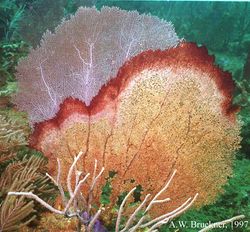
Two types of red band disease exist: RBD-1, which closely resembles black band disease, except that the bands are maroon in color and lacks the white specks; RBD-2 has cyanobacterial filaments that spread like a net on the surface. RBD leaves behind exposed coral skeleton. The pigment is caused by a different genus of cyanobacteria as the dominant microorganism, and the migration pattern across the surface of the colony also differs from BBD.[8]
White Band Disease
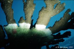
White band disease is a common coral disease detrimental to elkhorn and staghorn coral. [9] Affected corals have thick strips of bleaching occurring at the bottom of the branch that spread upwards. The bleaching is caused by the expulsion of symbiotic algae while tissue pulls away from the skeleton starting at the base. It is suspected that WBD is caused by a bacterial infection. [10] There are two types of the disease, White Band Disease 1 (WBD1) and White Band Disease 2 (WBD2). The spread of the disease must be observed in order to determine which type it is. In type 1 there is a strip of lysis (cell break down), and in type 2 the coral degradation eventually catches up to this line of lysis.
This is one of the only diseases known to cause major changes in the structural composition of reefs. This disease was first documented in 1979, and since that time it has destroyed up to 95% of the acropora genus corals in the Caribbean.[11]
White Plague Disease
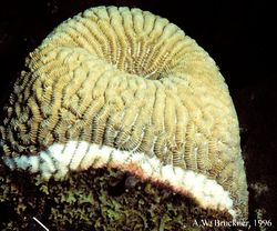
White plague disease is similar to the white band disease in that it causes portions of coral to bleach. However, the white plague disease affects a wider range of coral. A symptom of this disease is irregular lesions that leave white patches on coral. [12] White plague disease has a similar pattern to the WBD that the tissue begins to pull away from the bottom leaving a large line of bleached coral.
Yellow Blotch Disease
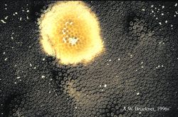
Yellow blotch disease causes sections of corals to become yellowed and translucent. Like many other coral diseases, the cause is unknown.[13] The disease begins at a central point anywhere on the coral and spreads outward. This is caused by a bacterial infection that causes yellowing of the tissue. As the infection spreads, the tissue is still alive, but eventually the tissue at the central point begins to die. This disease mostly affects large boulder star corals. Unlike most other diseases, recently bleached, white skeleton is rarely observed; typically the area behind the band is covered in filamentous and coralline algae.
Implications of Diseases in Reefs
Coral reefs are an important factor for communities, especially along the costal regions. Reefs are important for economies since they bring in money through tourism and the fishing industry. The weakening reef system may pose a threat to the countries that rely on this factor as a major component of their economy. Diseased reefs do not have the same tourist draw or fishing production as a healthy reef. Furthermore, reefs provide a barrier to protect the inland from large waves and surges. [14] The event of coral disease outbreak will have major negative implications. Therefore, more work and research should be focused on the causes in order to prevent further degradation of the reef ecosystem.
Coral disease hasn’t been thoroughly studied because it does present some challenges. First, it is hard to run experiments on sick coral that may have very short life spans and migrates across the coral. Second, there is an enormous variety between microbes and bacteria that cause disease. Lastly, spatial and temporal variation add an exponential amount of variability when trying to generalize results. We need are more marine microbiologists who can conduct research in the field and also understand the ecological and environmental effects on coral.
We must also consider that even though disease can very much occur naturally - humans can and do impose threats that may spread or worsen the toll disease takes on corals. Pollution and waste may be facilitators in spreading disease. Sea rise and temperature increase may increase the susceptibility of coral, and ocean acidification may weaken the corals’ immune systems. It is important that we do our part to protect the reef. By making small changes and encouraging sustainable behavior within the tourism industry, it can help the health of the reef ecosystem. Many of the chemicals used in sunscreen and unsustainable relaxation activities (golfing, boating, etc.) can cause added stress to corals, which make them more susceptible to diseases.
References
- ↑ http://coraldisease.org/
- ↑ “Coral Diseases” Reef Research Center. Retrieved 26 Feb. 2013 from <http://www.reef.crc.org.au/discover/coralreefs/Coraldisease.htm>
- ↑ 3.0 3.1 3.2 3.3 3.4 3.5 Field Guide to Western Atlantic Coral Diseases and Other Causes of Coral Mortality. http://www.masna.org/portals/0/NOAACoralDisease/index.htm
- ↑ “Major Reef-building Coral Diseases.” CoRIS - Coral Reef Information System. NOAA, 01-17-13. Web. 2-26-13. <http://coris.noaa.gov/about/diseases/#redband>
- ↑ Gochfield, Deborah, Julie Olson, and Marc Slattery. “Colony Versus Population Variation in Susceptibility and Resistance to Dark Spot Syndrome in the Caribbean Coral Siderastrea Siderea.” Diseases of Aquatic Organisms 69 (2006): 53–65. Inter-Research. Web. 02-26-13. <http://www.int-res.com/abstracts/dao/v69/n1/>
- ↑ http://coraldisease.org/diseases/12
- ↑ http://www.artificialreefs.org/Corals/diseasesfiles/Common%20Identified%20Coral%20Diseases.htm#BlackBand
- ↑ http://archive.rubicon-foundation.org/xmlui/handle/123456789/9047
- ↑ “Major Reef-building Coral Diseases.” CoRIS - Coral Reef Information System. NOAA, 01-17-13. Web. 2-26-13. <http://coris.noaa.gov/about/diseases/#red band>
- ↑ Aronson, Richard B. and Precht, William F. “White-band disease and the changing face of Caribbean coral reefs.” Hydrobiologia 460: 25-38
- ↑ Lentz JA, Blackburn JK, Curtis AJ (2011) Evaluating Patterns of a White-Band Disease (WBD) Outbreak in Acropora palmata Using Spatial Analysis: A Comparison of Transect and Colony Clustering. PLoS ONE 6(7): e21830. doi:10.1371/journal.pone.0021830
- ↑ “Major Reef-building Coral Diseases.” CoRIS - Coral Reef Information System. NOAA, 01-17-13. Web. 2-26-13. <http://coris.noaa.gov/about/diseases/#red band>
- ↑ Kellogg, C. A. “Montastraea cavernosa.” Photo. microbiology.usgs.gov 26 Feb. 2013. <http://microbiology.usgs.gov/image_gallery_plants_animals_montastraea_cavernosa.html>
- ↑ “Socioeconomic Impacts” Coral Reefs. Retrieved 26 Feb. 2013 from <http://www.reefresilience.org/Toolkit_Coral/C2c2_Socioecon.html>